Article Type: Case Report, Volume 3 Issue 1
*Corresponding author: Vartan Matossian
Santa Barbara Cottage Hospital Internal Medicine Residency Program, 400 West Pueblo Street, Santa Barbara, CA 93105, USA.
Email: vmatossi@sbch.org; vartanmatossian@gmail.com
Received: Dec 05, 2025 Accepted: Jan 06, 2026 Published: Jan 13, 2026
Citation: Matossian V, Carbone G, Lawlor C, Hosea S, Perrin M, et al. Leprosy unmasked by immunosuppression: A diagnostic pitfall with cutaneous sarcoidosis mimicry. Ann Case Rep Med Images. 2026; 3(1): 1061.
Copyright: Matossian V et al. © All rights are reserved
Background: Leprosy, also known as Hansen’s Disease, is a bacterial infection of the skin and peripheral nerves caused by Mycobacterium leprae [1]. Despite being uncommon in developed nations, leprosy remains a significant global health concern, with approximately 250,000 new cases diagnosed annually [2]. Cutaneous sarcoidosis is a granulomatous skin condition characterized by reddishbrown papules, plaques, or nodules that typically present on the face, trunk, or extremities. Cutaneous sarcoidosis can resemble leprosy due to similar clinical features like hypopigmented or infiltrated lesions, peripheral nerve involvement, and a chronic, indolent course.
Case presentation: A 65-year-old female with a past medical history of cutaneous sarcoidosis, aortic valve stenosis, remote history of Hodgkin’s lymphoma treated with radiation therapy, and hypothyroidism presented with one week of worsening facial and oral labial ulcers complicated by weeks of ongoing malaise and fatigue, neuropathy, as well as ongoing weight loss due to the inability to swallow. Notably, she had recurrent bleeding ulcers that had begun roughly eight months before presentation and were thought to be related to her immunotherapy treatment for sarcoidosis. However, these lesions had not resolved with numerous adjustments in her medications and immunotherapy. Remarkably, a biopsy of the lesions during her hospitalization revealed that her underlying diagnosis was leprosy.
Conclusion: Given this elusive clinical presentation, the physical exam and cutaneous biopsy were instrumental in pursuing the diagnosis despite the documented medical history. Importantly, images of the patient’s presentation were crucial for facilitating appropriate referrals and pursuing diagnostic tests, e.g., obtaining a biopsy and communicating with outpatient specialists.
Keywords: Sarcoidosis; Leprosy; Hansen’s disease; Mycobacterium leprae; Mycobacterial infection; Chronic infectious disease.
Leprosy, otherwise known as Hansen’s Disease, is a chronic granulomatous bacterial infection of the skin and peripheral nerves caused by Mycobacterium leprae [1]. The earliest documentation of leprosy dates back to about 2000 BCE in the ancient Sanskrit text Atharva Vida [1]. Though less uncommon in developed nations, 225 new cases of Leprosy are diagnosed in the United States annually [2]. Notably, the burden of disease is concentrated in a few countries, with India, Brazil, and Indonesia accounting for nearly 80% of cases [3].
There are two commonly known subtypes of Hansen’s disease: Pauci-Bacilliary (PB) which is also known as tuberculoid leprosy, and Multibacillary (MB) which is also known as lepromatous leprosy [4]. In addition, the Ridley-Jopling classification system identifies 5 subtypes of leprosy: Tuberculoid (TT), Borderline Tuberculoid (BT), Mid Borderline (MB), Borderline Lepromatous (BL), and Lepromatous Leprosy (LL) [5]. This classification system expands upon the aforementioned bipolar classification system to include histologic and immunologic features.
Skin lesions associated with leprosy may exhibit several morphologic features depending on the severity and type of disease. This leads to common misdiagnosis, especially in individuals with few risk factors. In PB leprosy, skin lesions often consist of a few well-defined macules that are anhidrotic, anesthetic, and hypopigmented to erythematous. Histopathology of these lesions is characterized by epithelioid granulomas with few organisms [6]. The lesions of Mycobacterial Leprosy (ML) may ulcerate and lead to deformity of facial structures. ML is associated with significant neurologic involvement, possibly even affecting vision, sense of smell, and speech [7,8].
The diagnosis of leprosy is made based on clinical findings, Slit Skin Smear (SSS), histopathology, and serologic testing [9]. The clinical findings of leprosy are broad, consisting of skin lesions of a variety of morphologies as described above and neurologic impairment of affected areas. Despite robust literature delineating the two major subtypes, little is understood about an individual’s predisposition to developing one form of the disease over another [10]. There are several studies identifying allelic genotypes and Single-Nucleotide Polymorphisms (SNPs) that are associated with both increased and decreased susceptibility to M. leprae, as well as whether an individual will develop PB or MB types [11]. Ultimately, the mechanism is likely multifactorial and poorly understood.
A 65-year-old female with a past medical history of cutaneous sarcoidosis, aortic valve stenosis, remote history of Hodgkin’s lymphoma treated with radiation therapy, and hypothyroidism presented to a California hospital with one week of worsening facial and oral labial ulcers complicated by weeks of ongoing malaise and fatigue, neuropathy, as well as ongoing weight loss due to the inability to swallow. The patient’s history was significant for progressive weakness of her extremities, leaving her in a cachectic state. Five days prior to presentation, the patient had been seen in the same emergency department with a similar rash and was discharged with triamcinolone. Symptoms only mildly improved before remitting, at which point the patient was seen by her rheumatologist via a telemedicine appointment and was instructed to re-present to the emergency department. Of note, two months prior to the hospitalization, she was treated for suspected shingles with acyclovir.
A physical exam was significant for numerous ulcers visible on the patient’s face, oral mucosa, and bilateral hands, knees, and feet. There were also mildly pink, erythematous nodules on her bilateral dorsal forearms consistent with the existing diagnosis of cutaneous sarcoidosis. Notably, the lesions of the ala of the nose and oral labia were ulcers with a hemorrhagic crust, but without surrounding erythema. On the brow bone and cheeks, there were a few shallow ulcers with surrounding scale. On the dorsal hands, the ulcers appeared punched-out. Ulcers of the palmar hands were shallow and the palmar surface of the hands appeared shiny and erythematous. There were also ulcers with hemorrhagic crust on the patient’s toes. The patient made the distinction that the bleeding ulcers were not consistent with any prior lesions related to her past diagnosis of cutaneous sarcoidosis.
Upon admission, the patient’s outpatient rheumatologist was contacted and reported that the diagnosis of cutaneous sarcoidosis was made by a dermatologist nearly four years prior, based on gross assessment of her skin lesions and tissue biopsy. The most recent bleeding ulcers had begun roughly eight months prior (though they were much less severe) and were thought to be a side effect of the mycophenolic acid she was prescribed for treatment of presumed cutaneous sarcoidosis. The lesions improved slightly when treatment was switched to etanercept one month later, though this was discontinued due to angioedema. Up until two months following this, the patient was on upadacitinib, but this too was discontinued at the time of shingles diagnosis (two months prior to admission), and hence the decision was made to start adalimumab. The most recent dose of adalimumab was noted to be three weeks prior to admission.
Lab studies were significant for a mild leukocytosis, thrombocytosis, and elevated C-reactive protein. Hemoglobin was moderately decreased, and calcium corrected for albumin was mildly elevated. Chest imaging was unrevealing for an acute process. While further investigation was pending, the patient was started on intravenous linezolid and acyclovir for the treatment of possible unresolved herpes zoster complicated by cellulitis.
Over the course of the hospitalization, the patient’s hoarse voice, dysphagia, and bleeding ulcers had improved. Meanwhile, an extensive infectious work-up resulted in negative findings for the following: Quantiferon gold, coccidioides antibody, histoplasmosis antibody, blastomyces antigen, varicella zoster virus PCR, and rapid plasma reagin. Coxsackie A immunoglobulin G was positive, while immunoglobulin M was negative. Coxsackie B 1-6 antibodies were also positive. Immunologic testing revealed elevated levels of immunoglobulins G and A, and decreased levels of CD3, CD4, and CD8 T-cells. Otherwise, there were normal levels of eosinophils, immunoglobulin E, tryptase, antinuclear antibody, complement factors 3, 3a, and 4a.
Given the unclear clinical picture, specialty consultations were pursued with colleagues in dermatology, rheumatology, ophthalmology, otolaryngology, and infectious disease. As a result, the decision was made to biopsy the lesions. Punch biopsies were performed at the bedside of the patient’s chin, forearm, and dorsum of hand lesions and sent out to pathology. Remarkably, the sample from the patient’s chin revealed suppurative and granulomatous dermatitis with erosions and ulcerations demonstrating numerous mycobacterial organisms and histiocytes with pale inclusions (noted as “globi”). Fite stain confirmed innumerable non-tuberculous mycobacteria. Confirmatory testing was pursued with samples being sent to both the Hansen’s laboratory in New Orleans and the University of Washington Medical Center, the latter of which validated the presence of Mycobacterium leprae. The patient was connected with the nearest Hansen’s disease clinic for optimal treatment and surveillance.
Leprosy is identified on clinical presentation utilizing three cardinal signs (though, even one of the following can lead the physician to strongly suspect the disease): loss of sensation in a hypopigmented or reddish skin patch, a thickened peripheral nerve with loss of sensation or weakness of the corresponding muscle, and a positive Acid Fast Bacillus (AFB) smear under slit skin smear examination. While the transmission of leprosy is not entirely understood, the most common route is humanto-human via prolonged exposure to respiratory droplets. Although rare, zoonotic transmission has been documented. Nine-banded armadillos are a natural reservoir for Mycobacterium leprae, and DNA from armadillos has been identified in isolates from infected humans [12]. The prolonged incubation period of up to 20 years makes the exact identification of the time of inoculation difficult. The fact that this patient had such obvious nasal and oral mucosal involvement leads us to believe the patient acquired the disease via respiratory tract transmission.
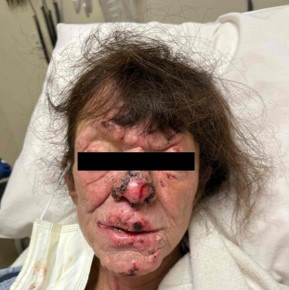
Figure 1: Leonine facies observed upon initial assessment.
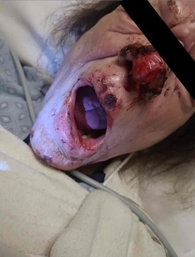
Figure 2: Ulcers and crusted lesions visible on face and mouth.
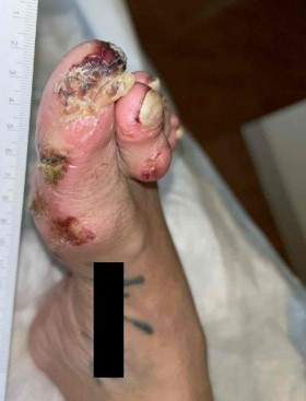
Figure 3: Ulcers and crusted lesions visible on right foot.

Figure 4: Ulcers and crusted lesions visible on right hand.
As far as exposures, the patient was born in California but reported extensive travel to endemic countries both as a young adult as well as up to five years before this presentation. International travel included camping in rural Baja California in the 1990’s, rural regions in the United Kingdom in the 2000’s where she was exposed to livestock, rural regions of France, India (including Goa, Mumbai, and Lavasa), rural regions of Guatemala, Greece, Jamaica, and Canada (nearby Toronto), and finally, another camping trip in rural Baja California in 2020.
This coincided with her initial cutaneous sarcoidosis diagnosis. While there is documentation of imported cases of leprosy from immigrants and refugees from endemic countries, we cannot definitively say if the patient acquired the infection overseas some years prior or while living in California [13].
Leprosy is a treatable chronic infection once identified by the practitioner. The initial form of the disease includes the undetermined form, which may or may not spontaneously resolve based on the patient’s innate cellular response to Mycobacterium leprae. As leprosy is far from being a common diagnosis, we believe the patient’s underlying disease was unveiled as a result of being appropriately treated with biologics over the course of eight months before admission with mycophenolate, infliximab, etanercept, adalimumab, and steroids for cutaneous sarcoidosis, which was considered the primary diagnosis at the time. During these eight months, her weakness and paresthesias began to progress, significantly impacting her activities of daily living. The adalimumab appears to have been the catalyst resulting in oral mucocutaneous and full-body rashes, as the patient was actively undergoing an adalimumab challenge within the preceding weeks before hospitalization. This can be explained by its Tumor Necrosis Factor (TNF) inhibiting nature, which may have reactivated or exacerbated the patient’s underlying leprosy. As TNF-α inhibitors, both etanercept and adalimumab impaired the body’s immune response to intracellular pathogens such as Mycobacterium leprae, leading to a reactivation or exacerbation of the patient’s underlying Hansen’s disease.
At the patient’s time of admission, she was initially treated with empiric linezolid for skin and soft tissue infection. Prior to identification of leprosy infection and proper therapeutic treatment, the patient began to improve on linezolid antibiotic monotherapy. While linezolid is not considered effective for treating M. leprae, it has been shown to have activity against other mycobacterial infections through inhibition of protein synthesis via binding to the 23S rRNA of the 50S ribosomal subunit [14]. While linezolid has not been studied in treating M. leprae, it is conceivable that it may have had a role in the patient’s improvement.
The current body of literature shows that TNF-α inhibitors, such as infliximab, etanercept, and adalimumab, have been correlated with the development or unmasking of latent leprosy infections. A meta-analysis of 20 studies revealed that among immunosuppressed patients with rheumatic diseases, 54.2% had been treated with biologics, and 95.2% of leprosy cases developed in this group [15]. Healthcare providers should consider leprosy in the differential diagnosis of patients presenting with skin lesions or neurological symptoms, especially those with a history of residing in endemic areas. Ongoing research and clinical consciousness are integral to balancing the benefits and risks associated with these treatments. Written informed consent was obtained by the patient prior to this publication.
Developing an expansive yet relevant differential diagnosis is a task that is undertaken by physicians daily. In the face of uncertainty, gathering key pieces of information and performing a thorough chart review are often essential. In cases such as the above, physical exam findings can be especially informative, given the presence of “leonine facies” that is usually described but best witnessed. Photographs were valuable in this case, as the experienced observer may incorporate broad differential diagnoses, such as leprosy, far sooner than without visual information, in order to then confirm the diagnosis via biopsy. A unique challenge in this example is questioning a prior diagnosis and how it may fit into the current clinical picture, especially given a seemingly discordant presentation and history. As well, a thorough social history to identify exposures, in this case pertaining to travel, may be highly informative and guide a physician down the correct path to diagnosis. Written informed consent was obtained from the patient for this publication.