Article Type: Case Report, Volume 2 Issue 1
*Corresponding author: Giancarlo Gismondo-Velardi
Department of Radiology, San Vincenzo Hospital, ASP Messina, Taormina, Italy.
Email: ggv84@hotmail.com
Received: April 19, 2025 Accepted: May 06, 2025 Published: May 13, 2025
Citation: Cipri C, Ferrari M, Maccarone R, Teti A, Gismondo Velardi G, et al. The role of CEUS - PMFI in the early diagnosis of middle hepatic vein thrombosis in a patient with multiple cryptogenic abscesses: A case report. Ann Case Rep Med Images. 2025; 2(1): 1023.
Copyright: Gismondo-Velardi G et al. © All rights are reserved
Hepatic Vein Thrombosis (HVT) is a rare but clinically relevant condition that can complicate hepatic pathologies and contribute to high morbidity. A prompt detection is crucial for therapeutic measures and prevent severe complications. We present a case of a 58-year-old male with middle hepatic vein thrombosis associated with cryptogenic hepatic abscesses. Contrast-Enhanced Ultrasound (CEUS), combined with Parametric Micro-Flow Imaging (PMFI), played a pivotal role in the early detection of the thrombosis.
Keywords: CEUS; Hepatic vein thrombosis; Parametric micro-flow imaging; Liver abscess; Vascular ultrasound; Early diagnosis.
Hepatic Vein Thrombosis (HVT) is a diagnostic challenge that can be life-threatening if not quickly identified. While most commonly associated with neoplastic liver disease or Budd-Chiari Syndrome [1], HVT may also occur in inflammatory settings, including hepatic abscesses [2,3]. In these cases, early detection of thrombosis becomes critical, as it can influence both prognosis and therapeutic strategy.
Contrast-enhanced Computed Tomography (CT) and Magnetic Resonance Imaging (MRI) have been considered standard modalities for detecting liver vascular complications. However, Contrast-Enhanced Ultrasound (CEUS) [4], especially when integrated with Parametric Micro-Flow Imaging (PMFI), offers the unique advantage of real-time vascular evaluation. CEUS-PMFI can detect subtle alterations in venous flow patterns, allowing for early visualization of thrombotic processes, especially in patients with suspected hepatic involvement but equivocal findings on standard ultrasound [5].
We describe the case of a patient with multiple cryptogenic liver abscesses in whom CEUS-PMFI enabled the early detection of middle hepatic vein thrombosis, allowing for prompt treatment initiation and favorable outcome.
A 58-year-old male presented to the emergency department with fever and right upper quadrant abdominal pain. Laboratory analysis revealed leukocytosis (WBC 25,950/µL, neutrophils 20,120/µL) and elevated C-reactive protein (347 mg/L). Blood cultures were negative.
First-line ultrasound examination showed multiple hepatic lesions. The largest lesion in segment V appeared iso-hypoechoic with heterogeneous and perilesional vascular signals. To better Jazzolino Hospital, ASP Vibo Valentia, Italy characterize the findings and evaluate potential vascular involvement, CEUS with PMFI was performed.
CEUS demonstrated a centripetal enhancement of the main lesion starting at 9 seconds post-contrast, with progressive filling up to 19 seconds. In the late arterial phase (25-30 s), early washout and central necrotic areas were evident. During the portal phase, additional lesions with peripheral enhancement and necrotic centers were observed.
CEUS-PMFI revealed an absence of flow within the middle hepatic vein and one collateral branch, indicating thrombosis. These findings were later confirmed by contrast-enhanced CT performed two days later. The patient was promptly started on broad-spectrum antibiotics and supportive care. Follow-up imaging showed clinical improvement and resolution of the thrombus.
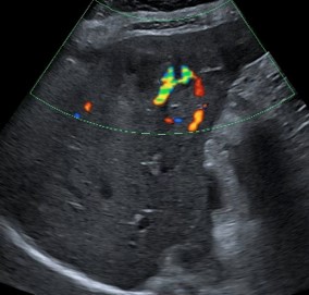
Figure 1: Ultrasound images showing largest lesion in segment V, appeared heterogeneous and iso-hypoechoic with prominent perilesional vascular signals on color Doppler.
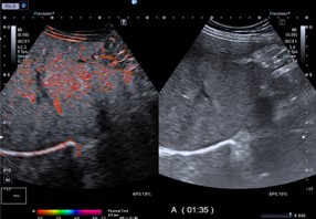
Figure 2: CEUS-PMFI post processed images demonstrate absence of flow in the middle hepatic vein.
Thrombosis of the portal or hepatic veins can occur as a complication of various underlying conditions, including hypercoagulable states, myeloproliferative disorders, inflammatory or neoplastic diseases, portal hypertension, and therapeutic interventions such as percutaneous or endoscopic injections. These potential risk factors should be taken into account when evaluating patients with focal liver lesions and suspected hepatic vein thrombosis.
PMFI is an advanced ultrasound technology that uses post-processed color-coded visuals to show wash-in time. It employs progressive frame accumulation, assigning each pixel a specific color according to the moment the contrast agent reaches the target region—defined as the point when pixel intensity exceeds a predetermined threshold. PMFI enables precise mapping of the time at which microbubbles enter and transit through the area of interest. This allows for comprehensive contrast-related information to be displayed in a single, intuitive image [6].
CEUS is routinarely used to characterize liver lesions. However, recent literature [5] underscores its role in evaluating hepatic venous flow, especially for patients needing rapid decisions, such as those with sepsis and suspected liver vascular involvement.
Hepatic abscesses often present with non-specific clinical symptoms such as fever, abdominal pain, and nausea. An early diagnosis represent a challenge in patients with vascular involvement, such as hepatic vein thrombosis. A prompt and correct diagnosis is mandatory to prevent complications such as hepatic infarction or sepsis [7,8].
This case demonstrates the potential of CEUS-PMFI in the early detection of hepatic vein thrombosis and often underdiagnosed. Color Doppler sonography plays a valuable role in characterizing thrombi. However, it is important to note that the sensitivity of this finding remains limited [9].
CEUS-PMFI enables high-resolution, motion-compensated imaging of low-flow vessels. In this case, early identification of hepatic vein thrombosis may have prevented further complications such as hepatic infarction or progression to Budd-Chiarilike physiology. Moreover, real-time CEUS evaluation allowed dynamic monitoring of lesion response during follow-up.
CEUS combined with PMFI enabled early diagnosis of hepatic vein thrombosis in a patient with multiple cryptogenic hepatic abscesses. CEUS-PMFI is useful not only for focal liver lesion characterization but also for evaluation of vascular involvement and permits a prompt and correct diagnosis which allows a correct therapy that improving the prognosis. The increasing use of CEUS-PMFI improves diagnostic accuracy of vascular involvement in hepatic pathologies.