Article Type: Short Report, Volume 1 Issue 1
*Corresponding author: Jessica Folk
Division of Emergency Medicine, North Shore University Health System, USA.
Clinical Assistant Professor, University of Chicago Pritzker School of Medicine, USA.
Email: jfolk@northshore.org
Received: Aug 18, 2024 Accepted: Sep 12, 2024 Published: Sep 19, 2024
Citation: Folk J, Mittal V, Haag A. Post traumatic splenic cyst. Ann Case Rep Med Images. 2024; 1(1): 1007
Copyright: Folk F et al. © All rights are reserved
A restrained 17-year-old male initially presented to the emergency department following a high-speed motor vehicle collision with no active complaints. Exam was reassuring nota ble only for a knee abrasion and patient was discharged home. The following day patient had some vague abdominal plate and the primary physician ordered an abdominal flat plate was obtained at that time (Figure 1). Labs completed on follow up one year later demonstrated hyperbilirubinemia (total bilirubin 2.1) and a repeat x-ray showed progressive hepatomegaly. The patient was referred to gastroenterology who noted significant abdominal distension and patient reported a 15-pound weight loss. Given concern for marked hepatomegaly, splenomegaly or situs inversus and patient was directly sent to the emergency department. CT imaging of the abdomen and pelvis was ob tained (Figure 2). A drain was placed with near resolution of the fluid collection (Figure 3).
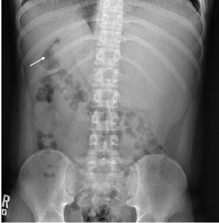
Figure 1: Abdominal flat plate x-ray showing a right upper quadrant stomach bubble (arrow) with left upper quadrant fullness suspect for situs inversus or other etiology.
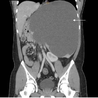
Figure 2: CT demonstrating intrasplenic fluid-filled mass (arrow) measuring up to 26 cm with significant mass effect on the left kidney, stomach and major vessels.
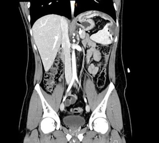
Figure 3: CT imaging one month after drain placement with residual fluid loculated and containing gas representing residual cyst (white arrow).
CT imaging revealed an intrasplenic fluid-filled mass with significant intra-abdominal mass effect (Figure 2). Interventional radiology percutaneously drained 6.5 L of sero-sanginuous fluid. With history and prior imaging this was suspect for a delayed traumatic cyst. One month later repeat CT imaging (Figure 3) demonstrated a redundant cyst wall. Patient underwent surgical deroofing of the cyst which unveiled multiple dense adhesions requiring complete splenectomy. Patient has received asplenic vaccinations. One year following his splenectomy he had mesenteric adenitis and was found to have chronic portal vein thrombosis with cavernous transformation and thrombocytosis, currently being monitored for possible anticoagulation given he is otherwise asymptomatic.
Initial flat plate x-ray on this patient following the motor vehicle collision was suspect for situs inversus with consideration of further imaging recommended based on presentation. In the setting of a possible solid abdominal organ injury, a contrastenhanced CT is recommended. It was unclear in documentation whether a FAST was performed which also could have aided in the diagnosis and prompted further imaging in a reassuring examination.
Splenic cysts, classified by tissue type and origin, have an incidence rate of 0.07% [1,3]. Traumatic subcapsular or intraparenchymal hematomas can result in post-traumatic splenic cysts [1,2,5]. Thirty to sixty percent of patients with splenic cysts are asymptomatic and do not require intervention [2,4]. Symptomatic patients, who often have left upper quadrant pain with nausea and vomiting due to gastric compression, will require treatment [2,4]. Eliciting a history of blunt abdominal trauma and identifying the splenic cyst on CT scan are critical in diagnosis [5]. As splenectomy patients hold a 0.2%-0.5% risk of significant infections techniques such as percutaneous drainage, marsupialization, splenic deroofing and complete cystectomy with partial splenectomy have been explored [2,4]. Although minimally invasive, case reports of percutaneous drainage have shown high recurrence rates, so complete cyst removal with partial or complete splenectomy should be considered for those with symptomatic splenic cysts [5].
The authors declare that they have no known competing financial interests or personal relationships that could have appeared to influence the work reported in this paper.
This case presents a pediatric patient who presented one year following a motor vehicle collision with delayed presentation of significant splenic cyst to the emergency department and importance of CT imaging with surgical consultation for management.
This project has not currently been presented at any meetings. No authors are sponsoring members.